
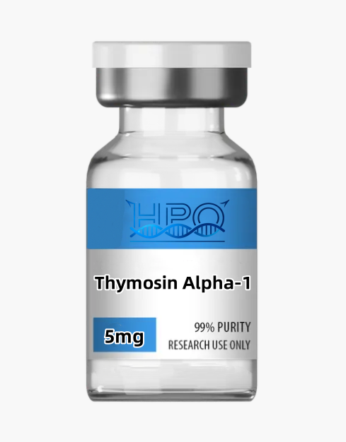
Thymosin Alpha-1
Price range: $80.00 through $160.00
Thymosin Alpha-1 research peptide (Tα1/thymalfasin) for dendritic-cell biology, TLR/MyD88-associated signaling, cytokine regulation, IDO research, and peptide analytics. Available in 5 or 10 mg × 10-vial configurations; laboratory research only—not for human or animal use.
DISCLAIMER:
Thymosin Alpha-1 Research Peptide: Comprehensive Laboratory Guide
Thymosin Alpha-1 research peptide (Tα1), also known in its synthetic form as thymalfasin, is a 28-residue, N-terminally acetylated acidic peptide used in laboratory studies of dendritic-cell biology, T-cell interactions, cytokine regulation, TLR/MyD88-associated signaling, p38 MAPK and NF-κB activity, and indoleamine 2,3-dioxygenase (IDO)-linked immune tolerance. It corresponds to the N-terminal region of prothymosin alpha. Published findings show that its effects vary with cell type, stimulus, receptor context, concentration, and experimental timing; therefore, it should be described as a pleiotropic immunoregulatory research tool rather than a universal “immune booster.”
This listing supplies Thymosin Alpha-1 exclusively for qualified nonclinical laboratory research. The available 5 mg × 10-vial and 10 mg × 10-vial configurations are catalog pack sizes, not experimental or clinical doses. The material is not a medicine, vaccine, diagnostic product, dietary supplement, cosmetic, compounded preparation, or product for human or animal administration. This page intentionally provides no injection, administration, self-experimentation, disease-treatment, or clinical dosing instructions.
Molecular and Product Overview
| Product name | Thymosin Alpha-1 research peptide |
|---|---|
| Common abbreviations | Tα1; Talpha1; thymosin α1 |
| Synthetic equivalent name | Thymalfasin |
| Peptide length | 28 amino-acid residues |
| Defined sequence | Ac-Ser-Asp-Ala-Ala-Val-Asp-Thr-Ser-Ser-Glu-Ile-Thr-Thr-Lys-Asp-Leu-Lys-Glu-Lys-Lys-Glu-Val-Val-Glu-Glu-Ala-Glu-Asn-OH |
| One-letter representation | [Ac]-SDAAVDTSSEITTKDLKEKKEVVEEAEN-OH |
| Molecular formula | C129H215N33O55 |
| Average molecular mass | Approximately 3108.3 g/mol |
| Registry information | CAS 62304-98-7 is commonly used for thymalfasin/Tα1; additional database mapping includes 69521-94-4. Verify the exact lot and reference standard. |
| PubChem CID | 16130571 |
| Listing configurations | 5 mg × 10 vials or 10 mg × 10 vials |
| Purity and form | Lot-specific; consult the certificate of analysis for chromatographic purity, peptide content, counterion, water, and other reported attributes |
| Research-use restriction | Laboratory research only; not for diagnosis, consumption, compounding, or human or animal use |
The formula and average mass above describe the defined peptide structure in a standardized chemical record. They do not by themselves describe every physical component in a supplied vial. Counterions, residual water, formulation components, and trace process-related substances can contribute to gross mass. Researchers should use the certificate for the exact lot received and independently qualify the material for the intended method rather than treating catalog fill mass or a single HPLC percentage as a complete quality statement.
Structural and Physicochemical Features
Thymosin Alpha-1 was originally isolated and sequenced as a small acidic thymic peptide. Its N-terminal acetyl group is part of the defined structure and is an important identity attribute. In mass analysis, the acetylated peptide is approximately 42 Da heavier than a corresponding des-acetyl form. A method intended to distinguish these species should use an appropriate intact-mass or peptide-mapping strategy rather than relying on chromatographic retention alone.
The sequence contains several aspartate and glutamate residues, which contribute to its acidic character, as well as multiple lysine residues. It contains no cysteine, so the defined peptide has no native intramolecular disulfide bond. It also contains no tryptophan or tyrosine and no phenylalanine; consequently, absorbance at 280 nm is not a reliable standalone concentration measurement. Peptide-bond detection near 214 nm, mass spectrometry, amino-acid analysis, or another validated quantitative method may be more appropriate depending on the study.
Solubility, adsorption, recovery, and solution stability can change with pH, ionic strength, buffer identity, concentration, container surface, temperature, and sample history. A universal solvent or universal solution shelf life should not be inferred from the sequence alone. Laboratories should perform small-scale, method-specific compatibility work and document actual recovery in the matrix and labware used for the assay.
Mechanisms Relevant to Laboratory Research
1. Pleiotropic and Context-Dependent Immunoregulation
Thymosin Alpha-1 has been associated with multiple immune-cell responses rather than one conclusively established, high-affinity receptor. Reported outcomes differ across dendritic cells, monocytes, T-cell co-cultures, microbial stimuli, and assay conditions. Therefore, mechanistic studies should avoid describing Tα1 as a selective receptor agonist. A stronger design compares multiple cellular contexts and uses pathway inhibition, genetic controls, time courses, and orthogonal readouts to determine which response is specific to the chosen system.
2. Dendritic-Cell Differentiation and Maturation
Human monocyte-derived dendritic-cell studies have reported changes in differentiation, maturation markers, and antigen-presentation-associated phenotypes after exposure to Tα1 under defined conditions. Dendritic cells integrate pattern-recognition signals and help coordinate adaptive immune responses, making them a central model for this peptide. Useful measurements can include morphology, viability, HLA class I and II expression, co-stimulatory markers, cytokine release, migration-associated markers, and the capacity to support T-cell responses.
3. TLR/MyD88-Associated Pathway Mapping
Research in fungus-pulsed dendritic cells linked Tα1-associated maturation and interleukin-12 production to MyD88-dependent Toll-like receptor signaling. Other work connected tolerance-related outcomes to TLR9 and type-I interferon receptor pathways. These observations demonstrate pathway dependence in particular models; they do not prove that Tα1 directly binds TLR2, TLR9, or another TLR as a conventional ligand. Direct binding, pathway requirement, and downstream modulation are separate questions and require different experiments.
4. p38 MAPK and NF-κB Signaling
Rapid activation of p38 mitogen-activated protein kinase and NF-κB has been reported in dendritic-cell research. These pathways can influence maturation, survival, transcription, and inflammatory mediator production. Time-resolved phosphoprotein analysis, nuclear translocation measurements, reporter assays, and selective pathway perturbation can help distinguish early signaling from later transcriptional consequences. Vehicle, stimulus-only, peptide-only, and inhibitor controls are necessary because many culture manipulations can independently activate these pathways.
5. Cytokine and Chemokine Modulation
Published cellular studies describe changes in mediators such as IL-12, IL-6, TNF-α, IL-8, IL-10, and type-I-interferon-associated responses. The direction and magnitude are not uniform. In human dendritic-cell systems, viral TLR3 or TLR7/8 contexts produced different response patterns from bacterial TLR2/4 or BCG-associated contexts. This stimulus dependence is scientifically important: researchers should report the agonist, donor, differentiation protocol, exposure sequence, sampling time, normalization method, and detection limits rather than generalizing one cytokine profile to all immune models.
6. IDO and Immune-Tolerance Research
A dendritic-cell study linked Tα1 to IDO induction, IL-10 production, and regulatory T-cell generation through TLR9- and type-I-interferon-receptor-dependent mechanisms. IDO catalyzes the first step in tryptophan degradation along the kynurenine pathway and can influence local immune regulation. Appropriate experiments may measure IDO transcript and protein, kynurenine-to-tryptophan ratios, dendritic-cell phenotype, and functional regulatory T-cell outcomes. Pathway inhibitors and receptor-deficient controls help separate correlation from requirement.
7. Dendritic-Cell and T-Cell Crosstalk
Tα1 research frequently examines how changes in antigen-presenting cells influence T-cell priming, polarization, expansion, or regulatory behavior. Co-culture systems can evaluate activation markers, proliferation, cytokines, transcription factors, and antigen-specific responses. Results depend heavily on donor variability, antigen quality, cell ratio, maturation state, culture duration, and background stimulation. A direct effect on T cells should not be inferred when only mixed-cell cultures were measured.
8. Peptide–Membrane and Biophysical Interactions
Biophysical investigations have considered how the peptide’s charge distribution and conformational flexibility may affect interactions with membrane-like environments. Such work can generate hypotheses about local concentration, orientation, uptake, or signaling access. However, membrane association is not equivalent to a defined cell-surface receptor interaction. Spectroscopy, membrane models, labeled-peptide imaging, uptake controls, and functional assays should be combined when testing these hypotheses.
Appropriate Research Applications
Dendritic-Cell Phenotyping
Thymosin Alpha-1 can be included in donor-derived or cell-line dendritic-cell workflows to investigate differentiation and maturation. Researchers may compare unstimulated cells, stimulus-only controls, Tα1 alone, and combined conditions across a predeclared time course. Flow cytometry, multiplex cytokine analysis, microscopy, and transcript profiling can provide complementary evidence. Batch effects, donor demographics, cell viability, and endotoxin controls should be documented.
Pattern-Recognition and Signal-Transduction Studies
Defined TLR agonists, MyD88 perturbation, type-I interferon receptor controls, kinase inhibitors, and reporter systems can be used to map pathway requirements. A concentration-response curve alone cannot establish direct target engagement. Mechanistic conclusions are stronger when genetic loss-of-function, rescue, phosphosignaling kinetics, and functional outputs converge on the same pathway.
Cytokine and Interferon-Response Profiling
Multiplex assays can examine whether Tα1 changes the magnitude or timing of responses to specified viral, fungal, or bacterial model stimuli. The purpose is to characterize experimental immune signaling, not to claim prevention or treatment of infection. Investigators should prespecify primary analytes, control for assay interference, use appropriate multiplicity correction, and report both absolute values and normalization procedures.
IDO, Kynurenine, and Regulatory T-Cell Models
Researchers can explore tolerance-associated biology by combining IDO expression, metabolite measurements, IL-10 analysis, and functional T-cell assays. Because regulatory T-cell phenotypes can be transient or marker-dependent, studies should use a defined marker panel and a functional suppression or equivalent validation assay where appropriate. Parallel viability and proliferation controls help prevent nonspecific cytotoxicity or growth arrest from being mistaken for immune regulation.
Peptide Identity and Impurity Method Development
The small, acetylated sequence is useful for developing LC-MS, MS/MS, reversed-phase HPLC, capillary electrophoresis, or related peptide methods. A stability-indicating method should distinguish intact Tα1 from des-acetyl, oxidized, deamidated, truncated, aggregated, or adsorption-depleted material where relevant. Retention time is method-specific; identity should be supported by a qualified reference and orthogonal evidence.
Comparative Immunoregulatory Peptide Research
Tα1 may be compared with thymic bioregulators, antimicrobial peptides, or inflammation-related research peptides to identify pathway-specific versus shared responses. Use matched matrices, verified identity, comparable active content, receptor-appropriate models, and the same analytical window. Results from one peptide or one cell system should not be extrapolated to clinical benefit.
Analytical and Quality-Control Framework
- Confirm provenance. Record supplier, lot, stated form, manufacturing date, storage history, vial condition, and all available documentation.
- Verify intact identity. Use LC-MS, Q-TOF, or another qualified intact-mass method and reconcile the result with the expected acetylated structure.
- Confirm sequence attributes. Apply MS/MS or peptide mapping when sequence, N-terminal acetylation, or positional identity is critical.
- Assess related substances. Use a suitable RP-HPLC or orthogonal separation method to evaluate impurities and degradation products.
- Establish peptide content. Distinguish chromatographic area purity from actual peptide content and total vial mass.
- Evaluate recovery. Test adsorption and matrix effects in the same tubes, plates, buffers, and concentration range used in the study.
- Qualify model-relevant contaminants. Measure endotoxin, bioburden, particulates, or synthesis residuals when required by the cellular or biochemical model.
- Retain raw data. Preserve chromatograms, spectra, integration settings, plate maps, acceptance criteria, and reserve material.
A reported HPLC area percentage is a method-dependent measurement and does not independently establish identity, peptide content, functional activity, sterility, endotoxin status, or suitability for a particular study. Likewise, an intact-mass match cannot exclude every positional isomer or low-level impurity. Fit-for-purpose qualification should combine chemical, physical, and functional evidence.
Experimental Design Controls
Good Tα1 research separates peptide effects from stimulus effects. Include vehicle, untreated cells, stimulus-only controls, Tα1-only controls, and a qualified positive or pathway-specific comparator. When investigating TLR-associated signaling, document the exact agonist, receptor context, cell source, donor, differentiation protocol, timing, and perturbation strategy. Include viability measurements and check for endotoxin because even low contamination can distort immune-cell data.
Use biological replicates that reflect donor or independent-culture variability, not only repeated wells from one preparation. Define exclusion criteria and normalization before analysis. When multiple cytokines or transcriptional endpoints are tested, apply an appropriate multiple-comparison plan. Report effect sizes and confidence intervals rather than relying only on thresholded significance.
Lot comparisons should use the same qualified reference, matrix, labware, and system-suitability criteria. If results change, investigate peptide identity, active content, impurities, adsorption, cell state, stimulus quality, instrument performance, and data processing before assigning the difference to biological potency.
Laboratory Handling and Stability
Qualified personnel should handle Thymosin Alpha-1 research material under a written institutional risk assessment. Use laboratory-appropriate personal protective equipment, avoid aerosol generation and accidental exposure, and keep the material segregated from food, medicines, clinical supplies, and personal-use devices. This page does not establish a hazard classification and does not replace lot-specific safety documentation.
Follow the lot label and certificate for storage. Peptide stability can depend on temperature, light, moisture, oxygen, pH, ionic strength, container surface, concentration, and repeated temperature cycling. For analytical solutions, use an institutionally approved, method-specific preparation based on measured solubility, recovery, and stability in the actual matrix. Low-binding labware and appropriately sized aliquots may be useful when adsorption or repeated handling affects recovery.
This page intentionally does not provide a reconstitution volume, administration route, injection concentration, clinical dose, or treatment protocol. Catalog vial size is not a recommendation for any experiment. Laboratories remain responsible for protocol approval, biosafety review, method validation, compliant storage, and disposal.
Frequently Asked Questions
1. What is Thymosin Alpha-1?
Thymosin Alpha-1 is a 28-residue, N-terminally acetylated acidic peptide corresponding to the N-terminal region of prothymosin alpha. The synthetic peptide is also called thymalfasin. Researchers use it to study dendritic-cell behavior, immune-cell crosstalk, cytokine regulation, TLR/MyD88-associated pathways, p38 MAPK/NF-κB signaling, IDO biology, and peptide analytical methods. This catalog material is supplied only as a nonclinical laboratory reagent.
2. Does Thymosin Alpha-1 have one confirmed receptor?
No single, specific high-affinity receptor has been conclusively established. Published studies report model-dependent involvement of TLR/MyD88, type-I interferon receptor, p38 MAPK, NF-κB, and other signaling components. Pathway dependence does not prove direct peptide binding to a TLR. Researchers should use direct binding methods, genetic perturbation, pathway inhibitors, and functional rescue designs when investigating target engagement.
3. Which assays are useful for Tα1 research?
Useful approaches include dendritic-cell differentiation and maturation panels, flow cytometry, cytokine multiplex assays, phosphoprotein analysis, NF-κB reporters, IDO expression, kynurenine measurements, T-cell co-culture assays, LC-MS, MS/MS, and RP-HPLC. Assay selection should match a defined hypothesis, with vehicle, stimulus, viability, endotoxin, receptor-pathway, and reference-material controls.
4. Does HPLC purity prove the material is suitable?
No. HPLC area percentage depends on the method, detector, integration, and sample preparation. It does not alone establish peptide identity, peptide content, N-terminal acetylation, biological activity, sterility, endotoxin status, or suitability for a specific cell model. Review the lot certificate and independently confirm the attributes required by the study.
5. Are 5 mg and 10 mg recommended doses?
No. They are catalog fill-size labels supplied in ten-vial configurations. They are not clinical doses, animal-study doses, recommended assay concentrations, or administration instructions. Researchers must define experimental concentrations from the assay range, qualified material, controls, institutional approvals, and applicable regulations.
6. Can Thymosin Alpha-1 be studied with other peptides?
A laboratory may compare or combine research reagents under an approved protocol, but each component should be tested alone as well as in combination. Use matched vehicle, concentration-response, time-course, viability, and pathway-specific controls. An apparent larger response does not by itself demonstrate synergy. This material must not be mixed or administered for personal, therapeutic, veterinary, or unapproved use.
7. Can this material be used to treat infection, cancer, or immune disorders?
No. Hanpro supplies this material solely for nonclinical laboratory research. It is not intended for diagnosis, prevention, treatment, vaccination, compounding, injection, ingestion, or human or animal administration. Findings from cell models, animal research, or regulated clinical literature cannot establish the identity, safety, or efficacy of a catalog research reagent.
Related Research Materials
- KPV — a tripeptide research material for inflammation-associated signaling models.
- LL-37 — an antimicrobial host-defense peptide for innate-immune research.
- ARA-290 — a tissue-protective receptor-pathway research peptide.
- BPC-157 — a research peptide used in tissue-response and experimental repair models.
- TB-500 — a thymosin beta-4-related research material for cell-migration studies.
- Humanin — a mitochondrial-derived peptide for cellular-stress research.
- VIP — a vasoactive intestinal peptide research material for receptor and immune-signaling studies.
- SS-31 — a mitochondria-targeted research peptide for cardiolipin and cellular-stress models.
- MOTS-c — a mitochondrial-derived peptide for metabolic-stress signaling research.
- NAD+ — a cofactor research material for redox and cellular-metabolism assays.
Quality Documentation
Researchers should request the certificate that applies to the exact lot received. Useful documentation may include intact-mass identity, chromatographic purity, stated peptide form, peptide content, counterion or salt information, residual water, and other lot-specific tests. Methods and specifications vary, so the receiving laboratory remains responsible for determining whether the evidence supports its intended use.
Do not infer sterile status, clinical grade, validated potency, endotoxin limits, or absence of a specific impurity unless lot documentation explicitly tests and supports that property. Retain receiving records, analytical raw data, and reserve material when traceability matters. Independent confirmation protects data integrity and downstream interpretation.
Selected Authoritative References
- PubChem. Thymosin alpha 1, CID 16130571. View the compound record.
- Goldstein AL, et al. Isolation and sequence characterization of thymosin alpha 1. View the PubMed record.
- Liu D, et al. Nα-acetylation and analytical characterization of thymosin alpha 1. View the PubMed record.
- Peng Y, et al. Thymosin alpha 1 and human monocyte-derived dendritic-cell differentiation. View the PubMed record.
- Romani L, et al. MyD88/TLR-associated signaling in fungus-pulsed dendritic cells. View the PubMed record.
- Romani L, et al. TLR9/type-I-interferon-dependent IDO and tolerance research. View the PubMed record.
- Giuliani C, et al. Stimulus-dependent effects in human dendritic-cell models. View the PubMed record.
- King R, Tuthill C. Mechanistic and membrane-interaction review of thymosin alpha 1. View the PubMed record.
Research-use disclaimer: This Thymosin Alpha-1 material is supplied exclusively for nonclinical laboratory research by qualified professionals. It is not a medicine, vaccine, compounded preparation, diagnostic device, food, dietary supplement, cosmetic, or veterinary product. It is not for injection, ingestion, diagnosis, treatment, prevention, patient use, or human or animal administration. Nothing on this page is medical advice. Investigators are responsible for biosafety assessment, ethics and legal review, method validation, safe disposal, and compliance with all applicable institutional and jurisdictional requirements.
| Specification | 5mg*10vials, 10mg*10vials |
|---|
Only logged in customers who have purchased this product may leave a review.
Reviews
There are no reviews yet.